Download the instruction video(15 MB) here, and use the PDF version (see below) for the details.
Download the PDF version (size is about 430 kB). In order to read the file, you need to have Adobe Acrobat Reader. (When you do not have Acrobat Reader, you can first download this software from the Adobe website.)
| Aspiration procedure | |
| Making glass slides | |
| Congo red staining and assessment | |
| Amyloid A protein, preparation | |
| Amyloid A protein, quantification | |
| Literature |
Aspiration of abdominal subcutaneous fat tissue is a simple outpatient procedure (1). It should be noticed, however, that it takes at least 10 - 15 minutes to avoid unnecessary pain and bruising and to get ample material. The patient should know that bruising might occur.
|
|
|
|
|
|
Equipment |
Practical |
Marking (1) |
Marking (2) |
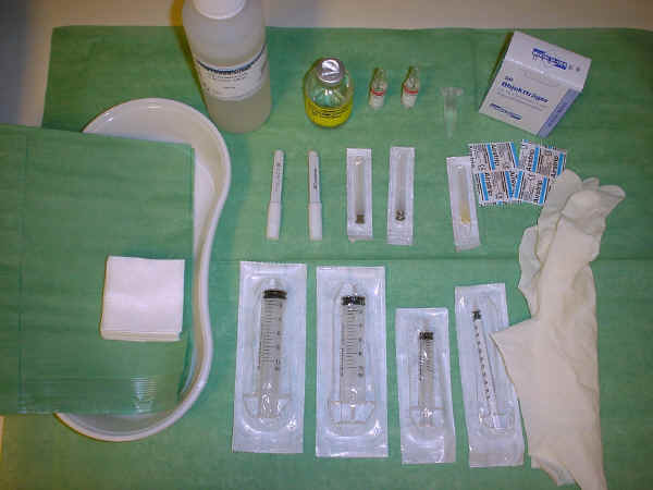
Chloorhexidine solution for skin cleaning, a 5 ml syringe connected to a 22 Gauge needle for lidocaine anaesthesia, two 10 ml syringes connected by a valve system to 16 Gauge needles for fat aspiration, small band-aids, gauzes, and protective gloves.
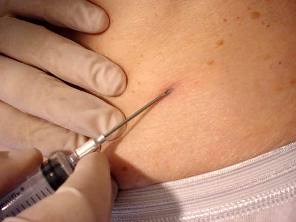
A syringe of 10 ml is connected by a valve system to a needle of 16 Gauge. After closing the valve the plunger is pulled out, fixed with squeezed fingers, and the cap of the lidocaine needle can be reused elegantly by positioning it upside-down inside the plunger ("Tarek's trick") to fix firmly the position of the plunger and thus maintain negative pressure in the syringe during aspiration. The skin of the patient is marked and cleansed (e.g. with chloorhexidine) at both sides of the umbilicus at about 7-10 cm distance. Skin and subcutaneous tissue (three directions, see below) are then anaesthetized with lidocaine (each side 2 ml=20 mg). Check first that the patient is not allergic to lidocaine. After inserting the needle beneath the skin the valve can be opened to start aspiration of fat tissue. The needle can be moved into three directions (Northeast, East, and Southeast) at the left side of the abdomen and mirror-wise at the right side. The aspiration procedure should be performed slowly and gently into each of the three directions, going to and fro with some rotation, and one should realise that it takes some time before the needle will be filled with fat tissue and the first fat can be seen passing the valve and entering the top of the syringe. This can be continued until enough fat tissue has been collected. After finishing the procedure press the puncture site for a while and cover the puncture site with a band-aid. Aim of the procedure is to obtain an adequate quantity for microscopic analysis (30 mg) and further at least 30 mg of fat tissue for immunochemical quantification of SAP and the specific amyloid proteins. Aspiration can be done at both sides of the umbilicus in order to obtain at least 60 mg of fat tissue.
|
|
|
|
|
|
Cleansing |
Lidocaine (1) |
Lidocaine (2) |
16 G needle |
|
|
|
|
|
|
Negative pressure |
A cap for ... |
... easy working |
Simple & easy |
|
|
|
|
|
|
Fat in citrate |
Without citrate |
Plaster |
The end |
 |
 |
||
| 50 mg in syringe | 50 mg of fat |
When you are finished and have collected enough fat tissue, the easiest solution for you is: Seal the syringes and ship them to Groningen for analysis at room temperature; see below for the shipment address.
After extracting the plunger, fat tissue can be collected from the syringe on an empty glass slide to separate fat tissue from accidentally obtained blood. At least four visible fragments of fat tissue (not fat droplets!) should be put on each of three glass slides (preferably with a frosted edge which can be used to write on it with a pencil). These fragments are crushed into a single layer by squeezing a second slide placed perpendicularly to the first ones. It is important to press in the middle of the glass slides to prevent breaking of glass. The resulting six smears are marked for identification, dried in the air at room temperature for one hour, and subsequently fixed with acetone for 10 minutes. After drying and fixation, all slides can be stored at room temperature until (shipped to a reference centre for) staining with Congo red and further study if positive for amyloid. Fat tissue should not be frozen before slides have been made: freezing of fresh and unfixed tissue may affect the quality of the tissue.
|
|
|
|
|
|
picking a lump |
lump on slide |
4 lumps / slide |
Perpendicular |
|
|
|
|
|
|
Pressing |
After pressing |
Separating |
To the lab |
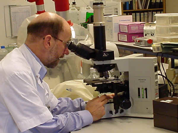
Staining with alkaline Congo red should be done according to the classic method described by Puchtler (2). The affinity of tissue for Congo red can be analysed by the apple-green birefringence in polarized light using a good microscope. In our institution we use the Olympus BX 50 microscope, 100 Watt, and we score the slides in a blinded way to the clinical data and semi-quantitatively: 0 (negative, no apple-green birefringence detectable), 1+ (minute, <1% of surface area), 2+ (little, between 1% and 10%), 3+ (moderate, between 10% and 60%), 4+ (abundant, >60%).
|
|
|||
|
Microscopy |
Below you can see an example of a fat aspiration stained with Congo red dye. This fat tissue has been collected in the outpatient clinics by using local anesthetics, some inches besides the umbilicus, just beneath the abdominal skin.
|
|
|
Ordinary light |
Polarised light |
As you can see above, amyloid can be recognized in the fat tissue as red stained deposits between the normal blue colored architecture of the tissue. When these red amyloid deposits are viewed in polarized light, the red areas have been changed into green, a characteristic feature of amyloid.
The remaining fat tissue can be stored in an 2 ml Eppendorf cup (at –20 C or
–80C) until shipment (at room temperature preferably within three days, but
ultimately within one week) to our hospital in Groningen, The Netherlands.
Address:
University Medical Center Groningen
Mr. Johan Bijzet, BSc
Lab. Rheumatology, T2.238 HPC EA41
Hanzeplein 1, 9713 GZ Groningen
The Netherlands
Tel: +31 50 361 3421
Fax: +31 50 361 3591
E-mail: j.bijzet@umcg.nl
|
|
|
|
|
|
Fat for extraction |
Plate |
ELISA procedure |
Software |
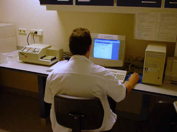
Before quantification, the amount of fat is weighed to get the so-called wet weight. It is then washed three times in a Tris buffer with calcium to remove possible remnants of blood still present. Then SAP is extracted by incubation with a Tris buffer with EDTA and the SAP concentration can be measured by ELISA. Subsequently the washed fat tissue is extracted in a solution of Tris and guanidine, centrifuged, and the supernatant of the fat tissue extract is collected. The total protein concentration and amyloid A protein concentration is measured by ELISA as has been described (3-5). Dutch reference values of patients without AA amyloidosis: < 12 ng/mg fat tissue or < 1.3 µg/mg protein. TTR and immunoglobulin kappa and lambda light chain concentrations can also be measured by ELISA, Western blot or Nephelometric methods.
1.
Westermark P. Diagnosis and
characterization of systemic amyloidosis by biopsy of subcutaneous abdominal fat
tissue. Internal Medicine Specialist 1984;5:154-60.
2. Puchtler H, Sweat F, Levine M. On the binding of Congo red by amyloid. J Histochem Cytochem 1962;10:355-63.
3. Hazenberg BPC, Limburg PC, Bijzet J, van Rijswijk MH. A quantitative method for detecting deposits of amyloid A protein in aspirated fat tissue of patients with arthritis. Ann Rheum Dis 1999; 58:96-102 > pdf >